

Diabetic retinopathy is a sight-threatening condition that complicates diabetes mellitus. Diabetes mellitus leads to many complications that can affect the whole body organs, but retinopathy is the most commonly diagnosed. It is the most common cause of blindness among diabetic patients and all adults under 65 years old.
Diabetes mellitus is a metabolic disease in which the blood glucose levels become high. This high glucose can cause damage throughout the whole body by damaging the blood vessels that supply the various organs. It can affect the kidneys, brain, nerves, and eyes. It may affect the eyes in many ways; it may cause retinopathy, cataract, and paralysis of ocular muscles.
As we said, diabetic retinopathy is the most commonly diagnosed and the leading cause of vision loss; thus, let’s discuss it.
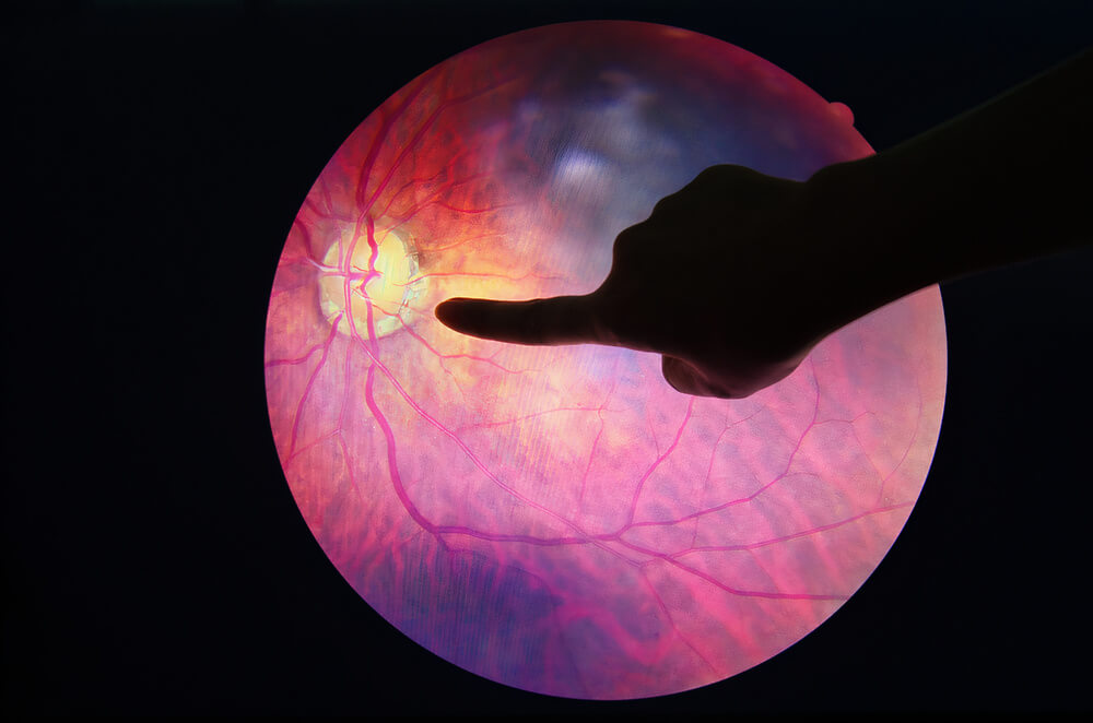
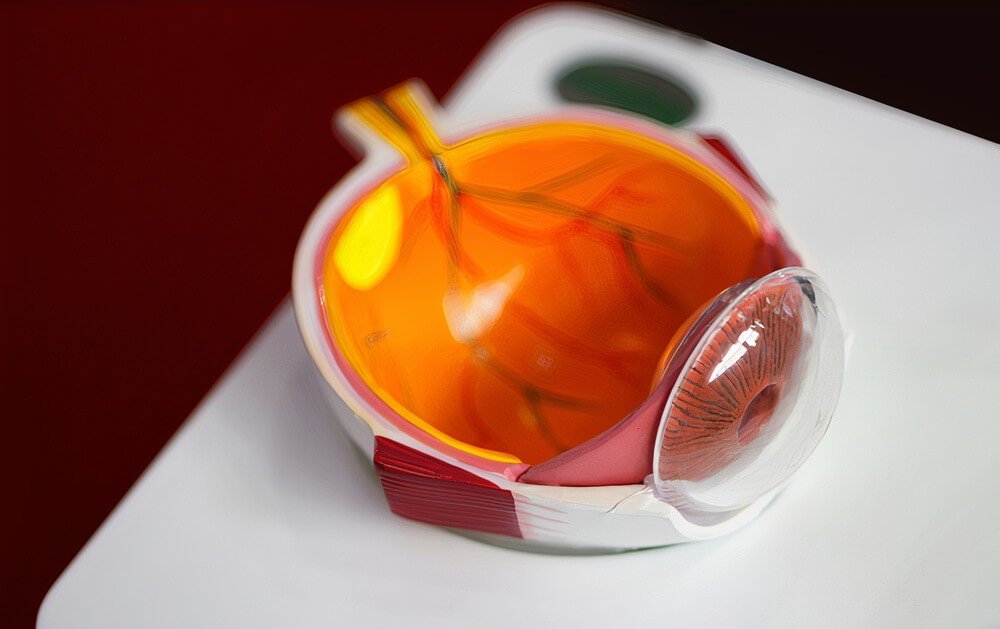
First, let’s know what the retina is. It is the light-sensitive layer of your eyes and lies a the back of your eye. It receives the light and changes it into signals that the optic nerve carries to the brain. Then, the brain translates these signals into the images that you see. The retina contains a part for sharp central vision, which is the macula.
In diabetic retinopathy, the high sugar damages and blocks the minute blood vessels that supply your retina. These blocked vessels will swell and leak fluids into the retina. Here, there are two problems: the swelling around the retina and the reduced blood supply (ischemia). Your body will respond to this by forming new vessels on the retina. But these new vessels are fragile and more liable to bleed.
Diabetic retinopathy takes several years to threaten your vision. But, you may not notice any symptoms until you reach an advanced stage. It develops gradually and ends in blindness. It also affects both eyes.
Any patient with diabetes can develop retinopathy, but the risk increases with the duration of diabetes. 20% of patients with type-1 DM will have retinal changes after ten years up to 90% after 20 years. About 20-30% of patients with type-2 DM have retinopathy at diagnosis. Also, prolonged poor control of diabetes increases the risk. We will discuss the other risk factors later.
Diagnosing retinopathy occurs by a detailed eye examination after taking a history of your symptoms. Your doctor may use some investigations to take images of your retina to see the abnormal findings and blood vessels in more detail.
Treating diabetic retinopathy depends on at which stage you are. In the early stages, you may not need any treatment, and it’s enough to manage your diabetes better and examine your eye regularly to assess the progression. In the advanced stage, your doctor will start active treatment to save your vision. The treatment depends on the problem in your retina, and we will discuss the options later.
Diabetic patients are at high risk, but they can take a role in preventing (or delaying) retinopathy and other complications. They can do this by following a good management plan for diabetes and regularly monitoring blood glucose. Also, regular eye exams (diabetic eye annual screening) help pick up the disease early and prevent (or reduce) its effect on vision.
Now, let’s dig deeper into the details of this condition -that is common among diabetic patients and impacts their lives-. We will answer the following questions:
-
- Who is at risk of diabetic retinopathy?
- How does this disease progress, and what are its symptoms and complications?
- How can your doctor diagnose retinopathy and assess its severity?
- How can your doctor treat you, and what are the available treatments?
- Can you protect yourself from vision loss by this disease, and how can you do this?
Are you at risk of diabetic retinopathy?

Any patient with diabetes mellitus can develop retinopathy, either type-1, type-2, or gestational (diabetes related to pregnancy). But, some risk factors may put some patients at a higher risk and require additional care, as follows:
The duration of diabetes:
The longer you have diabetes, the more the risk is for retinopathy.
Poor control of blood glucose levels:
Persistent high glucose levels enhance the harmful effect of glucose on blood vessels, which increases any retinal damage.
Hypertension (high blood pressure):
Like diabetes, hypertension has a damaging effect on blood vessels and causes hypertensive retinal changes that worsen the damage of diabetes. Most patients with diabetes develop hypertension, which aggravates its complications.
Hyperlipidemia (high cholesterol level):
It causes more damage to the blood vessels and fastens their damage by diabetes. It’s also a risk factor for hypertension and its complications.
Pregnancy:
The risk of retinopathy is higher for a diabetic woman who got pregnant and a woman who developed gestational diabetes. Thus, the pregnant woman should do a comprehensive eye examination and additional eye exams during pregnancy. Pregnancy also increases the risk of all complications of diabetes.
Smoking:
Smoking increases the risk of all diabetes complications, not only retinopathy. It also increases the risk of hypertension and its complications.
Race:
Hispanics and African Americans are at a higher risk.
Your doctor will consider these risk factors when managing retinopathy to reduce its progression. Also, you should know and avoid these risk factors to reduce the risk of retinopathy.
How does this disease progress? (Stages of retinopathy)

Diabetic retinopathy develops gradually over time. This disease begins with high glucose, which damages the blood vessels throughout the body, including the retina. This damage occurs in stages, as follows:
1) Stage 1 (Background retinopathy):
Small retinal vessels are blocked and swell; thus, tiny bulges appear on the retina (microaneurysms). These swollen vessels leak fluid to the retina.
At this stage, you don’t have any symptoms. But you are at a high risk of vision problems later. You also don’t need active treatment, but you should control your glucose level more effectively to prevent progression.
2) Stage 2 (Pre-proliferative retinopathy):
More damage to the retinal vessels occurs, and your doctor sees more changes in the retina, such as bleeding into it. Also, these damaged vessels can’t supply the retina with adequate blood (ischemia).
At this stage, there is a much higher risk of vision affection. Also, you will need more frequent eye screening exams (every 3, 6, or 9 months) to monitor your eyes.
The previous two stages represent the non-proliferative type of retinopathy. In this type, your condition progresses from mild to severe with more damage to blood vessels.
3) Stage 3 (proliferative retinopathy):
It is the most advanced and severe type of diabetic retinopathy. In this type, your body responds to retinal ischemia by stimulating the growth of new abnormal vessels on the retina. But, these new vessels are fragile and more likely to bleed and leak fluid easily into the vitreous (jelly-like fluid fills in front of the retina). According to the amount of this bleeding, you may see a few dark floaters (if slight) or lose your vision (if much).
Also, scar tissue forms with the new vessels formation, which may pull on the retina and causes a retinal detachment. Also, these new vessels interfere with the normal flow of fluid from the eyes, which increases the intraocular pressure. High intraocular pressure cause glaucoma and destroys the optic nerve.
At this stage, you are at risk of losing your sight. You can’t restore what you lost; thus, you need active treatment to save the remaining of your vision.
4) Diabetic maculopathy:
The macula lies central to the retina; it is responsible for the central vision. Sometimes the damage of retinal blood vessels affects the macula, leading to fluid leakage (macular edema) and (or) macular ischemia. Macular edema requires treatment to avoid permanent vision loss because it is the most common cause of vision loss among patients with diabetes.
What are the symptoms and complications of this disease?

As we said, diabetic retinopathy develops gradually through stages. The early stages are asymptomatic. You have a clear vision, but the disease progresses. Thus, attend the regular screening appointments to catch this condition early.
You will notice changes in your vision when the disease reaches its advanced stages. These changes include:
-
- You may see floaters (spots or dark shapes floating in your visual field).
- You may have a blurry vision that may sometimes change from blurry to clear.
- You may have poor night vision and have difficulty seeing in the dark.
- You may feel hard to read and drive (impaired central vision).
- Impaired color vision: you see faded or washed out colors.
- You may see holes, blank, or dark areas in your vision field.
- You may feel pain or see redness in your eyes.
- Finally, your may lose your vision. Vision loss may also be sudden and total.
Diabetic retinopathy usually affects both eyes.
Complications:


Diabetic retinopathy doesn’t affect the vision alone; it causes other problems that worsen the condition, such as:
Vitreous hemorrhage:
As we said, the newly formed fragile vessels on the retia may easily bleed to the vitreous, and the amount of bleeding may make floaters in your visual field (if slight) or block it (if much). Vitreous hemorrhage alone doesn’t cause permanent blindness because the blood clears from the eyes spontaneously. Your vision may return clear again if there is no retinal damage.
Retinal detachment
The newly formed vessels may also stimulate scar formation that pulls the retina and detach it. Retinal detachment may cause you to see light flashes or floating spots, or you may lose your vision.
Glaucoma
The newly formed vessels may extend to the iris (in the anterior chamber of the eyes), interfere with the normal fluid drainage out of the eyes, and increase the intraocular pressure, causing glaucoma. This condition may damage the optic nerve and cause permanent blindness.
Diabetic macular edema
Macula gives you a sharp and focused central vision that helps you read, drive, and recognize faces. Macular edema is the most common cause of vision loss among these patients. 50% of patients with diabetic retinopathy develop macular edema at any stage but more likely in the later stages. Vision loss due to macular edema is usually irreversible.
Blindness
You may lose your sight due to any of the above complications or a combination of them. The risk is higher with poor management.
How can your doctor diagnose diabetic retinopathy?

At first, your doctor will hear your history to know your vision difficulties and how you control your diabetes. He also will ask you if you have other health problems that may affect your sight, like hypertension.
Then, your doctor will do a comprehensive eye exam, as follows:
Dilated eye exam
Your doctor will place eye drops to dilate your pupil to allow him to see inside your eyes. Then, he will use a specific magnifying lens (ophthalmoscope) to look closely at the retina and detect any abnormalities. These eye drops may cause blurred near vision till they wear off (some hours later).
Visual acuity test
By the eye chart, your doctor will evaluate your central vision.
Tonometry
It’s the standard test to measure intraocular pressure (IOP).
Your doctor may need additional tests to assess the severity of your condition, such as:
Fluorescein angiography:
After dilating your eyes with specific drops, your doctor will inject fluorescein dye into your arm vein. This dye circulates with your bloodstream and reaches the retinal blood vessels. Via a special camera, your doctor can take images of your retina and detect the leaking, damaged, and blocked retinal vessels. This information helps your doctor assess the condition and represents guidance for the treatment.
OCT (optical coherence tomography):
This technique uses light waves to make a detailed cross-sectional image for the retina without dyes. These images reveal the retinal thickness, which helps your doctor determine swellings and how much fluid has leaked. OCT exam also detects any optic nerve damage. Your doctor may also use this exam to monitor the treatment’s effectiveness.
How can your doctor manage diabetic retinopathy?


Management of diabetic retinopathy depends on which type you have and its severity. The management plan aims to slow or stop the disease progression. But, whatever the type of retinopathy you have, managing your blood sugar is crucial.
Managing your diabetes:
In the early stages, managing your diabetes prevents developing vision problems. While in the late stages, it helps stop the disease progression.
Early diabetic retinopathy (mild or moderate non-proliferative type):
In these early stages, you may not need any treatment. Just conservative watchful waiting is enough. Your eye doctor (ophthalmologist) will do a frequent comprehensive eye exam (every 2 or 4 months) to determine when you will need treatment. Also, managing your diabetes is vital in these stages to avoid developing vision problems.
Advanced diabetic retinopathy:
Proliferative diabetic retinopathy or maculopathy requires immediate treatment because both conditions threaten your sight. Your doctor will decide the treatment, depending on the specific problem in your retina. The treatment options include:
1- Eye injections
Your doctor may inject anti-VEGF (vascular endothelial growth factor) medications into your eyes to stop growing new vessels on the retina and reduce fluid leakage. These drugs can slow down the disease progression and may improve your vision.
Your doctor injects these drugs under cover of local anesthetic drops. You may need to repeat these injections or use them with other options, like the laser.
2- Laser treatment (photocoagulation)
Your doctor may use the laser to shrink the abnormal vessels and seal the leaks points, which stops (or slows) the fluid accumulation within the retina. This treatment stabilizes your condition and prevents worsening, but it can’t improve your sight.
Your doctor will do this procedure after using local anesthetic drops. It takes around 20 to 40 minutes and doesn’t need a hospital stay. Also, you may need to repeat it more than once.
3- Eye surgery (vitrectomy)
If you have vitreous hemorrhage or retinal detachment, your doctor can do this surgery. The surgeon creates a small incision in your eye. Then, through it, he removes the vitreous (or some of it), the leaked blood, and the scar tissue (that pulls on the retina).
Your doctor will do this procedure under local anesthesia. You can go home on the same or the next day after surgery, but you need to wear an eye patch for a few days.
Your doctor will discuss these treatment options with you, tell you the benefits and side effects, then choose the most effective one for your case and determine the frequency. But, even after treatment, you need a regular eye exam because you may need additional treatment at some point.
Finally, how can you prevent diabetic retinopathy and its complications?

Diabetes is a lifelong disease, and you can’t always avoid diabetic retinopathy. But, it doesn’t always lead to vision loss. You can take an active role in reducing its risk or -at least- preventing severe vision loss by doing the following:
Manage your diabetes
Keep a healthy diet, healthy lifestyle, and regular exercise. Take your medications and insulin as your doctor prescribed and follow his recommendations.
Monitor your blood sugar
Check and record your blood glucose, especially under stress, like illness. Ask your doctor how you can do this and when you should worry about your glucose level. Ask him about the HB-A1C test that reflects the glucose control over the previous three months. Most patients should have HB-A1C less than 7.
Stop smoking
Smoking increases the risk of retinopathy and all other complications of diabetes. Stopping is necessary.
Monitor your blood pressure and cholesterol levels, and keep them under control.
Attend your diabetic eye screening appointments to catch retinopathy in its early stages before causing severe damage.
Seek early medical advice.
Call your eye doctor when you feel vision changes, such as hazy, blurry, or spotty vision.
Diabetes puts your sight at risk, but you can reduce this risk and save your eyesight by following an effective management plan for your diabetes.