
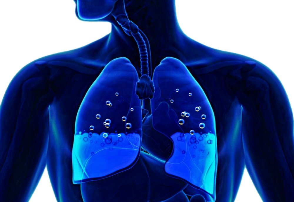
Pulmonary edema is a clinical condition in which excessive fluid accumulates within the air spaces of the lungs. This may occur suddenly or over a prolonged interval of time. Generally speaking, symptoms that appear suddenly are usually severe, while those that appear more slowing are more-mild, at least initially.
Symptoms of Pulmonary Edema
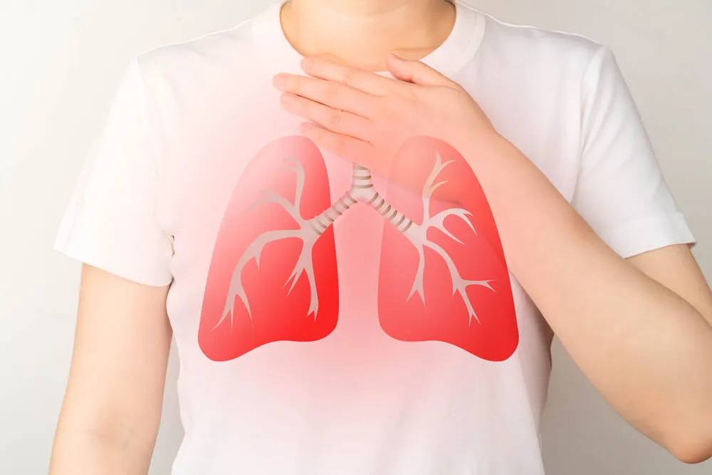
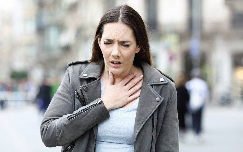
1. Difficult breathing
A healthy person at rest is usually not aware of their breathing. Respiration happens more or less automatically and is not given much thought. Heavy exercise causes breathing to become unpleasantly perceptible, but this is both normal and temporary.
Difficult breathing is the most common symptom of pulmonary edema. Depending on the cause of pulmonary edema, this symptom may develop suddenly or worsen slowly. Breathing may at first become difficult when performing strenuous activities, then during normal routine activities, then lastly at rest. Patients often become tired much more quickly than usual, and effortless actions may become burdensome over time.
In some cases, breathing may become difficult, particularly when the patient is lying flat on their back. These patients may relieve their symptoms by sleeping on multiple pillows to elevate the head and upper body. Other patients may complain of repeatedly waking up in the middle of the night, feeling out of breath and needing to sit up or to stand by an open window to recover. These two variants are caused by redistribution of blood from the legs to the chest when lying down.
Patients may describe this symptom using a variety of expressions, such as being uncomfortably aware of their breathing, feeling of chest tightness, feeling of choking or drowning, or being unable to take in enough air.
The presence of fluids in the air sacs of the lung impairs its ability to deliver oxygen to the blood, and this makes breathing difficult.
2. Cough

One of the most alarming symptoms of pulmonary edema is coughing up blood. The patient usually does not cough frank blood but rather frothy sputum streaked or tinged with blood, appearing pink in color.
3. Wheezing

Some patients with pulmonary edema may hear a high-pitched whistling sound while breathing. This is termed a wheeze. Wheezes occur due to narrowing of the airways.
If you imagine the air flowing at a high velocity through a narrow tube, you’ll get some idea of the mechanism of wheezing.
4. Swelling

Pulmonary edema is often caused by left-sided heart failure, where the blood inside the lungs cannot pass to the heart through the pulmonary veins due to dysfunction of the pumping mechanism of the left ventricle, and thus the lung’s blood vessels become congested, leading to the escape of fluids into the air sacs. In patients with co-existing right-sided heart failure, swelling may occur in various sites.
The right side of the heart receives blood returning from all over the body (except for the lungs) through large veins called the superior vena cava and inferior vena cava. This blood has already delivered oxygen and nutrients to the organs of the body and is now returning to the heart to be re-oxygenated. If the right side of the heart is diseased, blood will accumulate inside the right atrium and ventricle, which make up the right side of the heart. Eventually, blood coming from all over the body will not be able to enter the heart, so it remains within the veins of the body, and thereafter this trapped blood may partially pass out of the blood vessels and accumulate between the cells of the body, similar to what happens in the pathogenesis of pulmonary edema. Thus, the patient may experience abnormal swelling of one or more of the following sites: The feet, ankles, legs, lower back, abdomen, hands, or around the eyes.
When a swollen area is pressed on by a finger, the finger leaves a temporary indentation or “pit,” which disappears in a few minutes. These patients may sometimes have a pressure sensation or pain in the upper right abdomen due to a concurrent swelling of the liver. The swelling may be associated with some rather rapid weight gain due to fluid accumulation in the body.
5. Anxiety

Abnormal difficulty in breathing and coughing up pinkish sputum often brings about feelings of anxiety, restlessness, and dread in pulmonary edema patients.
6. Sweating


Excessive sweating and cold, clammy, abnormally pale skin are possible associations in pulmonary edema.
7. Palpitations

We are normally unaware of the heartbeats within our chest. When the number of heart-beats per minute increases or decreases significantly, or the heart pumps blood more forcibly, we become aware of our heartbeats. These unpleasantly perceptible beats are called palpitations.
8. Bluish lips


The impaired ability of the lungs to deliver sufficient oxygen to the blood due to the fluid present in the air sacs decreases the total amount of oxygen in the blood. This deoxygenated blood adds a bluish tinge to the lips, and sometimes to other sites such as the ears and the nail beds as well. In dark-skinned individuals, this manifestation may be less apparent.
9. Mental effects


The decrease in the amount of oxygen reaching the brain in pulmonary edema patients has a number of consequences, including an impaired ability to concentrate, decreased attentiveness, poor judgment, easy and rapid fatigability, apathy, and incoordination of movements.
10. Symptoms of the precipitating illness

Pulmonary edema has many causes, each of which has their own specific signs and symptoms. For example, renal failure patients have diminished urine output throughout the day, while patients suffering from pneumonia may have fever and chills. Convulsions, chest pain, and hives are other possible associated symptoms.
» Now, Let’s discuss pulmonary edema causes.
What causes pulmonary edema?
1. Heart failure


The most common cause of pulmonary edema is congestive heart failure. Typically, the heart pumps blood from the lungs into the aorta, the biggest blood vessel in the body.
A diseased heart, however, may not pump the blood efficiently. Thus, with each heartbeat, more and more blood accumulates in the heart’s left ventricle. This excessive volume causes the pressure to increase in the pulmonary veins, the vessels that transport blood from the lungs to the heart. Eventually, the pressure becomes so excessive that a lot of blood stays in the lung vessels, and then moves into the air spaces.
The causes of congestive heart failure are many, from high blood pressure to heart attacks, and even coronary artery disease.
2. Acute Respiratory Distress Syndrome

Acute respiratory distress syndrome or ARDS occurs mainly in critically-ill patients in the ICU. The most common cause of this condition is a severe infection of the bloodstream. A lung infection or pneumonia is another cause. Damage to the delicate membranes around the air sacs allows fluid to come in, making it difficult for oxygen to enter the blood.
3. Renal failure

In normal circumstances, the kidneys get rid of fluids that the body no longer needs and eliminate them into the urine. If the kidney function fails due to a severe infection, massive blood loss, a blood clot, or any other cause, the fluid will start to build up in the lungs, and breathing becomes difficult.
4. Seizures


A dangerous condition caused by electrical and chemical dysfunction in the brain. Seizures are defined by loss of consciousness and uncontrollable movement of the whole body or a part of it. Occasionally, a seizure may be followed by pulmonary edema. This is likely due to the excessive activation of the sympathetic nervous system, the fight-or-flight mechanism of the body.
5. Aspiration

Choking occurs when a foreign material enters the airway, and further damage is normally prevented by coughing the offending substance out of the airways. In patients under anesthesia or with neurological disease, where the cough reflex is suppressed, there is a risk of gastric juice accidentally getting into the lungs. The strong acidity damages the lung tissue and allows fluid to enter.
6. Re-expansion

Rapid re-expansion of the lung is another cause of pulmonary edema. This can happen after treatment of lung collapse, which is often due to excessive fluid or air in the sac that envelopes the lungs, known as the pleura, which usually contains only a tiny amount of lubricating fluid.
7. Airway obstruction


During the normal process of breathing, negative pressure is generated in the lungs to suck the air in. This pressure should not exceed a particular range. When something blocks the airways, such as a piece of food or a coin, the lungs respond by putting in more effort to inhale air, resulting in increased negative pressure. Eventually, the air sacs and their thin membranes cannot stay intact and become ruptured and flooded.
8. Inhalation injury


Inhaling toxic or very hot gases can injure the lungs, causing chemical irritation or thermal damage respectively. Depending on the cause, there may also be external burns or a hoarse voice.
9. Trauma

A direct blow to the chest, such as during a car crash, may injure the lung’s smallest vessels, the capillaries, causing the blood inside them to leak into the pulmonary tissues.
» Now, Let’s discuss treatment of pulmonary edema.
Treatment of Pulmonary Edema
The first three essential steps of resuscitation are often referred to by using the easy-to-memorize acronym “ABC”, which stands for Airway, Breathing & Circulation. Making sure the ABCs are normalized is the first step in many other clinical conditions.
1. Airway
The physician first needs to make sure that the airway is open, and relieve any obstruction that may be present. Obstruction of the airway is not only caused by foreign objects, but may also be caused by the tongue or aspirated gastric juices. The patient is first encouraged to cough in order to expel the obstructing object.
Techniques used to clear the airway include the Heimlich maneuver, where the healthcare personnel stands behind the patient, puts his arms around the patient’s torso, and performs multiple inwards and upwards abdominal thrusts alternating with back slaps to force the foreign body out of the airway. In children under 12 months, the child is placed head down on the doctor’s leg.
Another method of securing the airway is tracheal intubation, where a tube is placed in the trachea to ensure its patency. More invasive procedures include the tracheostomy, in which a surgical incision is made in the neck and a tube is inserted within, connecting the trachea directly to the outside world. This is a last resort operation to relieve airway obstruction.
2. Breathing

Oxygen administration is crucial in the treatment of pulmonary edema, since the main pathology features a decrease in blood oxygenation. Methods of oxygen delivery include the face mask, nasal cannula, and mechanical ventilation.
3. Circulation

The most common cause of pulmonary edema is congestive heart failure, where an overworked or diseased heart is unable to pump blood efficiently, resulting in the accumulation of blood in the pulmonary vessels, which is then filtered into the lung’s air sacs. Thus, a variety of drugs are indicated to decrease the workload of the heart in these patients.
4. Diuretics


The kidneys get rid of excess fluids throughout the day by pumping unneeded minerals into tiny tubules that pass urine out of the body. Water follows the mineral atoms by the process of osmosis.
Furosemide, a potent diuretic, increases the volume of urine by increasing the amount of minerals in urine, specifically sodium, potassium, and chloride. With less fluids in the body, the heart has less work to do, and thus can pump more effectively.
5. Nitroglycerin
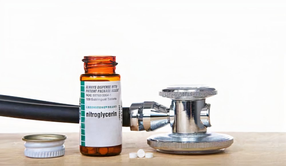

A very popular drug used for many heart conditions. Nitroglycerin is classified as a “Preload reducer”, which means it dilates the veins of the body, thus decreasing the amount of blood that goes to the heart. With less blood to become filled with, the heart function improves.
6. ACE inhibitors
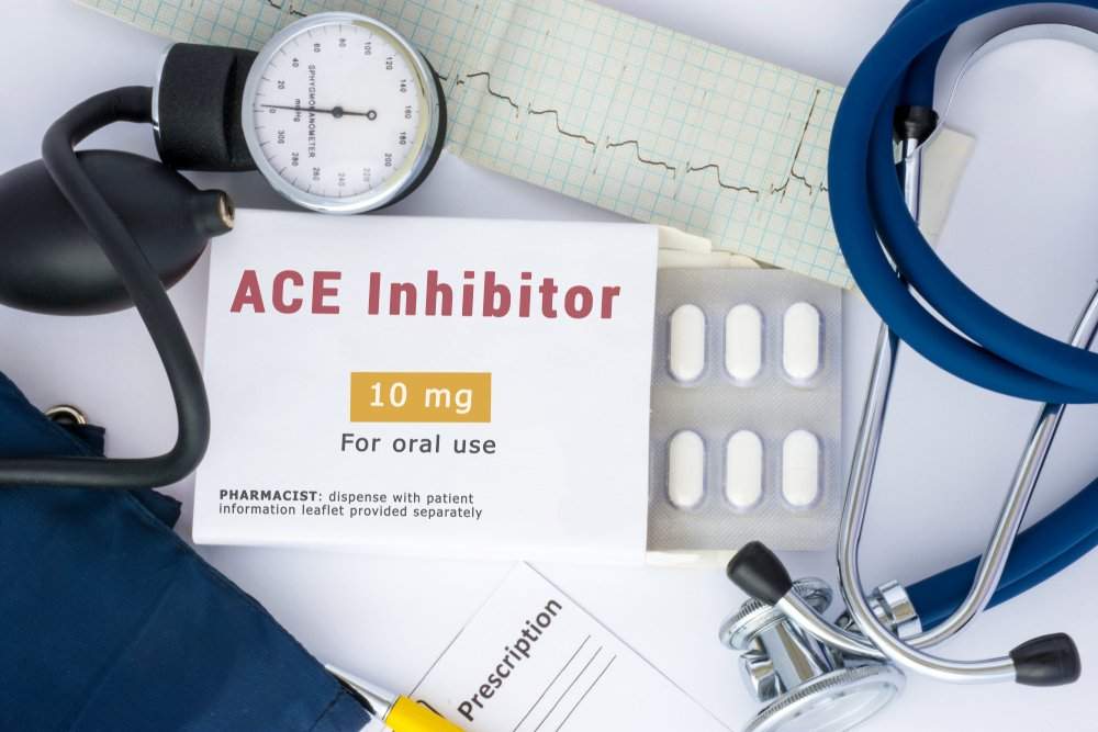
The Renin-Angiotensin-Aldosterone System (RAAS) acts to maintain blood pressure, blood volume, and systemic vascular resistance (how narrow the blood vessels are). Like many physiological mechanisms in the body, the “RAAS” may harm rather than heal the body.
A complex series of chemical reactions is initiated when the kidneys detect insufficient blood flow. Of particular interest is the formation of a potent peptide, Angiotensin II, by the enzyme Angiotensin Converting Enzyme (ACE). Angiotensin II is the main component of RAAS. It causes the blood vessels to narrow (Vasoconstriction), which makes it harder for the heart to pump against this increased vascular resistance. It also releases Aldosterone, a hormone that alters the constitution of urine, increasing the amount of fluids in the body. This eventually overloads the heart.
ACE inhibitors, such as Captopril, prevent the formation of Angiotensin II by blocking the action of Angiotensin Converting Enzyme (ACE). Therefore, these drugs decrease blood pressure, blood volume, and systemic vascular resistance. They are henceforth called “Afterload reducers”. Thus, the workload of the heart is decreased.
7. Angiotensin II receptor blockers


Patients who cannot tolerate ACE inhibitors can be given Angiotensin II receptor blockers (ARBs), which produce the same outcome without as many side effects.
Throughout the complex mechanism of action of ACE inhibitors, the normal rate of breakdown of a peptide called Bradykinin is decreased. Accumulation of Bradykinin causes a persistent dry cough as long as the medication is being taken in some patients. This is not seen with ARBs. An example of an Angiotensin II receptor blocker is Valsartan.
8. Inotropic drugs

Inotropes are a class of drugs that effectively strengthen heart contractions.
1) Dopamine:
A natural molecule synthesized in the body. Dopamine can be administered to increase cardiac contractility, dilate the blood vessels, and increase urine output, making the heart’s workload lighter.
2) Dobutamine:
This drug acts to increase the strength of heart contractions, increase the heart rate, and dilate the blood vessels of the body, making the heart’s job easier.
3) Milrinone:
A drug that belongs to the class of Phosphodiesterase inhibitors. Milrinone acts to increase cardiac contractility, decrease preload (less blood goes to the heart), and decrease afterload (Less systemic vascular resistance).
4) Levosimendan:
Calcium is the main molecule involved in muscle contraction, not just in the heart but throughout the body. Levosimendan makes the muscle layer of the heart, the myocardium, more sensitive to calcium ions. Thus, the strength of heart contractions is increased without needing excessive calcium entry, which would be dangerous. Levosimendan also causes dilatation of the blood vessels (Vasodilation) which makes cardiac pumping easier.
9. Sodium Nitroprusside

10. Intra-Aortic Balloon Pumping
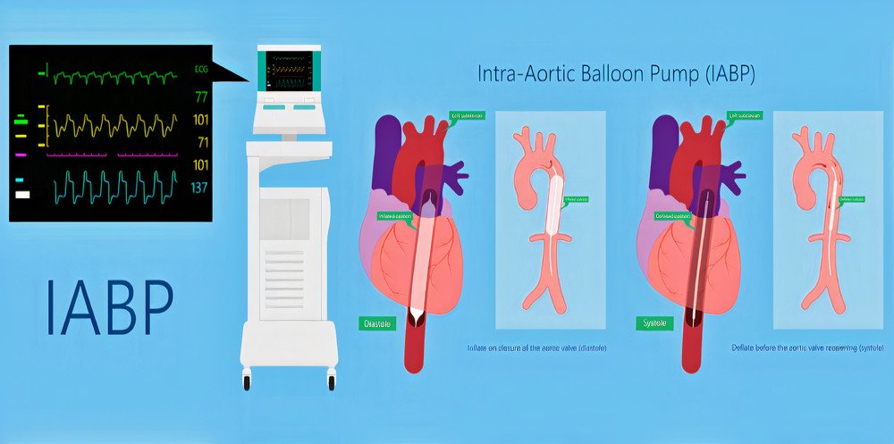

A temporary surgical procedure which is sometimes life-saving. A surgical incision is made in the leg, and the device is inserted into the femoral artery. The cylindrical balloon is advanced through the vascular system until it reaches the aorta, the body’s biggest blood vessel, which originates directly from the heart.
When the heart contracts, sending blood forcefully into the aorta, the helium-filled balloon is deflated, effectively creating a vacuum, which decreases the vascular resistance and facilitates blood flow.
When the heart relaxes, the balloon is inflated, sending some of the blood back to the coronary arteries near the origin of the aorta. The coronary arteries supply the heart itself with blood, containing oxygen and nutrients. The increased myocardial blood supply helps maintaining effective cardiac contractions.
The inflation and deflation of the balloon is controlled by a computer to match the rhythm of the heart. The balloon is filled with helium because helium is less likely than air to cause an embolism in case of ruptures. Radiography is used to make sure that the balloon is placed properly. The intra-aortic balloon pump is typically left in place for a couple of days.
› Treatment of the underlying cause
Depending on the disease that caused the pulmonary edema, specific treatments should be administered. For example, patients with heart valve disease may need surgical correction or a prosthetic heart valve.