

What is hyperopia?
Hyperopia is a common refractive error of vision; it is also known as farsightedness. Hyperopia is a frequent problem in the general population and occurs in children and adults. In hyperopia, the light rays focus just behind the retina not on the retinal surface as in emmetropia. Patients with hyperopia refraction error can see far objects clearly, where the near objects appear blurry.
Accommodation system of the eye tries to overcome this condition by:
-
- Increasing the corneal curvature
- Increasing the optical power of the eye lens
So, in patients with mild refraction, the accommodation will compensate for the refraction error and the patient will has no symptoms. In cases of accommodation dysfunction, the refraction error will manifest, and the symptoms and signs of the patient will appear according to the level of his refraction error.
In the humans, the newly born babies’ vision is usually on the hyperopia side and shifts slowly to be emmetrope or even myope in a few years.
Because hyperopia is a common complaint; proper assessment, diagnosis, and even preventing its complications is a public health responsibility to prevent its impact on the whole society. Genetics and family inheritance have a profound impact on hyperopia incidence. So, a widespread screening for hyperopia in young children especially in children with a family history is mandatory for early detection and treatment of hyperopia. This may help to prevent visual impairments, and other associated comorbidities.
Prevalence of hyperopia
Hyperopia is a common refractive error around the world. Castagno et al. leaded a meta-analysis in 2014 about the prevalence of hyperopia in school age children. They founded that at age 6-year-old 8.4% of the children had hyperopia, while in children from nine- to fourteen-year-old (9-14) the prevalence ranged from 2% to 3%. In fifteen-year-old group, the prevalence was 1%. In the same study they founded that prevalence of hyperopia was more in white children especially in rural communities.
In a review by T. Hornoch, showed that hyperopia prevalence could be affected by ethnicity and positive family history of hyperopia or accommodation dysfunctions.
Another study by Hashemi et al. to revise the prevalence of refractive errors around the world founded that the prevalence of hyperopia in adults and children was at its highest in the United States of America.
Lin et al. founded that the prevalence of moderate degree of hyperopia at six-year-old children was 13.2%, while at twelve- year- old children moderate hyperopia prevalence was 5%, with a higher prevalence in white Americans in comparison to other ethnic groups.
In a study to evaluate the prevalence of hyperopia and its geographical distribution in Southern India, Dandona et al. founded that, females with hyperopia were more than males in two age groups:
-
- Younger than fifteen-year-old
- Older than thirty-year-old
In a study by Christine L Allison to study the refraction error in polish immigrants in Chicago. She founded that hyperopia is the most prevalent refraction errors in the group of the study and is more common in the people ages forty-five-year-old and older.
In Cameroon, a study by Eballe et al. founded that, hyperopia is the most prevalent refraction error in children ages from six-year-old to fifteen-year-old.
➠ Now, let’s discuss causes of hyperopia
Causes of hyperopia

There are different causes for hyperopia, as there are multiple types of hyperopia with different etiologies. Hyperopia is classified according to its etiology into:
1- Axial hyperopia
It is the most common type of hyperopia; it is a simple hyperopia. Axial hyperopia usually results from shortening of the axial length of the eye. In this type genetics plays significant role in the developing of simple hyperopia. A study by Strang et al. founded that shortening of the axial length of the eye by 1mm leads to +6 diopters refractive error.
2- Index hyperopia
It occurs in patients with diabetes or in old age. In this type there is a change in the power of the crystalline length, which causes progression to hyperopia.
3- Curvature hyperopia
In this type, hyperopia may result from flattening of the cornea, or flattening of the crystalline lens, or both.
4- Positional hyperopia
In positional hyperopia, it results from
-
- changing the position of the crystalline lens in the eye
- absence of the crystalline lens due to genetic or acquired diseases
- Post- traumatic
- Post-surgical loss of the crystalline lens
5- Functional hyperopia
Malfunction of accommodations which may appear since birth causes functional hyperopia. Other causes of accommodation disfunction includes using cycloplegic drugs, or in cases of third nerve palsy.
6- Miscellaneous
A few conditions can be associated with hyperopia such as microphthalmos, Anophthalmus, and aniridia. There are multiple diseases and environmental factors that could be linked to hyperopia, from these factors:
-
- Family history
- Excessive smoking during pregnancy
- Diabetes mellitus
- Optic nerve edema
- Retinal edema
- Heimler syndrome
Stages of hyperopia
According to American optometric association, hyperopia can be classified into multiple stages or degree
-
- Low hyperopia: in this degree the patient has +2 diopters or less
- Moderate hyperopia: in this degree the patient has +2.25 to +5 diopters
- High hyperopia: the patient has +5 diopters or more
Symptoms of hyperopia


The symptom of hyperopia depends on the age of the patient and the degree of hyperopia. The age of the patient at presentation is particularly important
- In young children, the child can not address his own complaints
- The accommodation effects may not be able to overcome high myopia degree in immature age
The degree of myopia has impact on the symptoms; on small refraction errors the accommodation can correct this error without the patient even notices, while in high errors the accommodation cannot compensate hence the patient may has clear symptoms. According to patient presentation, the patients of hyperopia can be classified into two categories:
1) Asymptomatic
In which the patient has a good ciliary muscle tone, and a good effective accommodation. This combination can overcome small degree of hyperopia without any notice from the patient.
2) Symptomatic
In this category the patient has a variety of symptoms or complaints such as
- Eye deviation: The parents usually notice this symptom. the deviation could be unilateral, or bilateral eye deviation, in this case the deviation could be alternative, or simultaneous. In a study by Lambo et al.founded that in young patient with hyperopia, the most common eye deviation was accommodative estropia, which is inward deviation of the eye. The prevalence of accommodation estropia in USA is near 1-2% of the population.
- Eye strain: it is also known as asthenopia, it is because of accommodation system working hard to correct the refraction error of hyperopia. The asthenopia is usually accompanied with other symptoms as headache, photophobia, lacrimation, and redness of the eye. The asthenopia can be triggered by prolonged sessions of near work activities.
- Diminished near vision: if the hyperopia is fully corrected with the effects of accommodations, after prolonged time, the patient may complaints of asthenopia and decrease in near object vision. If the hyperopia is higher more than the effect of accommodation to correct, the patient usually complains of diminished of near and far vision.
- Pseudo-myopia: it is a sudden attack of temporary blurry vision in adolescents and young adults with uncorrected hyperopia. This symptom may be due to prolonged use of accommodations.
- Recurrent conjunctivitis: refraction errors cause frequent rubbing of the eye, which could be a source of infection to the eye.
- Early-onset presbyopia: it is a gradual deterioration of the eye ability to visualize the near objects. In normal populations, presbyopia is part of the aging process; the normal person starts to experience it in early or mid-forties. In hyperopia, due to the deterioration of the accommodation power, the presbyopia may appear earlier than in emmetrope.
Clinically, hyperopia is classified into three main categories: simple, pathological, and functional
- Simple hyperopia:it is due to disturbance of the normal functions and structures of the eye
- Pathological hyperopia:it occurs following trauma, surgical, or diseases in the eye
- Functional hyperopia:it occurs in cases of accommodation fails or dysfunction
Diagnosis of hyperopia

The evaluation and diagnosis of the patient with hyperopia aims to assess his clinical conditions, identify related conditions and possible co-morbidities, and correct of the patient refraction errors. Clinical evaluation of hyperopia assesses different aspect of the eye structurally and functionally.
– Assessing the patient visual acuity
The visual acuity of the patient depends on multiple factors such as
-
- accommodation power to fully correct the patient hyperopia or not
- age of the patient on the time of the evaluation
- internal structure of the eye especially crystalline lens and the posterior chambre of the eye
so visual acuity in children may not be affected due to full accommodation effect, and there is no associated eye condition e.g., cataract, glaucoma, and other retinal diseases. In high degree of hyperopia distant vision could also be affected. Associated amblyopia would also compromise the visual acuity of the patient.
– Diffuse illumination by slit lamp
The slit lamp could give a good picture about the eye structures. In hyperopia the cornea and eyeball may be smaller. Examination of the anterior chambre could reveal shallow anterior segment. The slit lamp is used with a gonio lens to examine the iridocorneal angle. The gonioscopy is a mandatory for all cases of hyperopia to exclude closure angle in patients with hyperopia. Cataract should be excluded in
-
- old age
- patients with diabetes
– Fundus examination
Fundoscopy is an important part of the clinical evaluation of hyperopia patients to rule out some associated retinal diseases such as pseudopopulist or pseudo papilledema.
– Cover test
This test is usually helpful in children with long term hyperopia without correction. Uncorrected hyperopia for long time may lead to accommodation squint.
Differential diagnosis of hyperopia


There are multiple conditions that could be interlapped or wrongfully diagnosed as hyperopia and should be kept in mind while diagnosing hyperopia such as:
-
- ptosis which may give a false picture of a small eye
- retinal edema
- nanophthalmous, in which the patient has normal structure of the eye, but the eye is smaller in size than normal eye
- microphthalmos, the patient’s eye is smaller in size with a structural abnormality
- Posterior microphthalmos
- Tumor mass in the posterior chambre of the eye
- Posterior scleritis
Treatment of hyperopia

Treatment of hyperopia focus on correction of the refraction errors, halting the development of amblyopia and prevention of strabismus, recurrent conjunctivitis, and its complications. Treatment measures includes
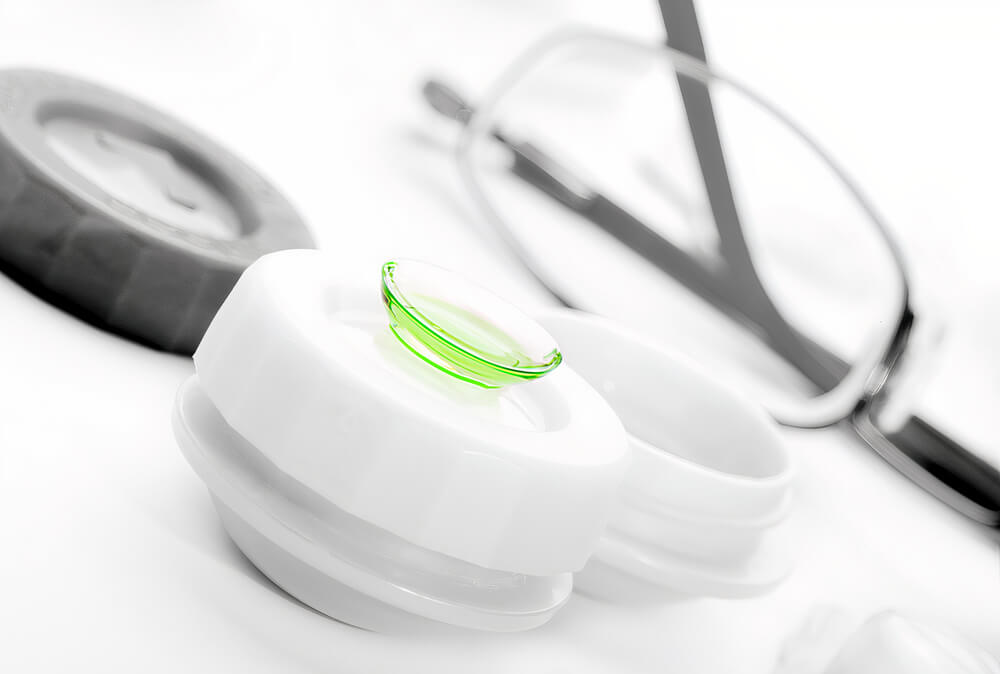
1- Non-surgical correction of hyperopia
Corrective lenses are the first choice in treatment of refractive errors. In hyperopia, if not associated with other diseases, the diminished visual acuity could be corrected by eyeglasses. To correct hyperopia, biconvex lenses are prescribed to help focusing the light rays on the retina. There are some important rules to follow when prescribing eyeglasses such as:
-
- In children with total hyperopia, the degree of hyperopia is determined by retinoscopy with complete cycloplegia
- In symptomatic hyperopia patient, especially in children and young adults, a full correction to the refraction error is mandatory
- In small errors e.g., +1 diopters or less, no correction is usually needed unless the patient is symptomatic
- Young children -before school age- can tolerate full cycloplegic correction, but should be closely monitored, and may be tarped later
- Older children may not be comfortable with the full correction of hyperopia. Correction can be increased gradually
- Children with hyperopia should be rechecked every six months
- Astigmatism should be fully corrected
- Accommodative squint needed a full cycloplegic correction of hyperopia
- If the patient developed amblyopia or lazy eye, a full correction with occlusion therapy is required
The American academy of ophthalmology (AAO) guidelines
prescribing eyeglasses in young children with hyperopia depends on multiple key factors. These factors include:
-
- Isometropia:it means both eyes have similar refractive errors
- Anisometropia:it means there are non-similar refractive errors in both eyes
- Prescence or absence of strabismus
In cases of isometropia, the eyeglasses are prescribed if the child has hyperopia of +3.5 diopters or more, in young children -below 2-year-old- is prescribed an eyeglass in cases of hyperopia of +4.5 diopters.
In cases of hyperopic isometric patient with accommodative squint, an eyeglass is prescribed when the refraction is +1.5 or +2 diopters.
In cases of anisometropia without strabismus, the patient should be prescribed an eyeglass in cases of
-
- In young children with hyperopia of +2 diopters
- In children from two-year-old to four-year-old with hyperopia of +1.5 diopters
The contact lenses are another option for optical correction, the use of the contact lenses is usually due to
-
- Aesthetic replacement to the eyeglasses
- If there were hyperopia in only one eye
- Anisometropia with high difference of the refractive errors between the two eyes
2- Surgical correction of hyperopia
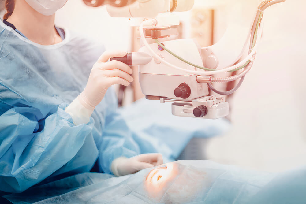
The surgical procedures can provide permanent correction of hyperopia, especially for patients with high refraction errors. Before any operation, the patient must have:
-
- The patient should maintain stable refraction error for at least three years before the surgery
- Slit lamp examination to rule out conditions like dry eye or conjunctivitis
- Full evaluation of the patient refractive errors using manual and cycloplegic techniques
- Evaluating corneal thickness
- Measuring intraocular pressure of the patient
- Fundus examination of the eye
- Evaluation of eye pupil size in light and dark conditions
Surgical procedures to correct hyperopia include:
A) Thermal laser keratoplasty
It is a non-contact laser thermokeratoplasty, which is indicated for hyperopia cases with refraction errors from +1 diopters to +2.5 diopters. In this procedure the laser causes dwindling of eight spots on the cornea, eventually this may lead to increase curvature of the cornea.
In a study by John C Hill, he founded that this procedure is slow in refractive errors correction, and do not treat the associated astigmatism.
B) Hyperopic photorefraction keratectomy (PRK)
It is a safe procedure for mild to moderate cases of hyperopia, it can treat a refraction error up to +7.5 diopters. An excimer laser is used to increase the curvature of the cornea. This procedure has a few disadvantages such as:
-
- It can cause dry eye
- Delayed healing of the epithelium
C) Hyperopic laser assisted in situ keratomileusis (LASIK)
Usually used to treat hyperopia patient with reflection error ranges from +1 diopters to +4 diopters and up to +6 diopters. Before LASIK surgery, the patient refractive errors must be stable for a considerable time. Contraindications of LASIK surgery includes:
-
- Chronic eye diseases
- Chronic corneal diseases
- Unstable refractions
- Dry eye
- Chronic pain syndrome
Although LASIK is mostly a safe operation, but a few complications may occur. From these complications
-
- Refraction deterioration
- Post-operative dry eye
- Halo vision
- Difficulty in night vision especially while driving
- Decrease corneal sensation
- Epithelial ingrowth
D) Hyperopic laser subepithelial keratomileusis (LASEK)
In this procedure, they use 20% ethyl alcohol for approximately sixty seconds, then the epithelium of the cornea is separated easily, and an excimer laser is used to perform the stromal ablation. In this procedure flap related conditions are much less in comparison to LASIK procedures, but the patent may experience more post-operative complications as:
-
- Post-operative pain
- Slow healing
This gives LASIK surgery a little bit advantage over LASEK
E) Conductive keratoplasty
This procedure for mild to moderate hyperopia. It is a non-invasive intervention, where low hyperopia could be corrected by using radiofrequency to increase the corneal curvature.
F) Phakic intraocular lens (IOL)
In this procedure, a new intra-ocular lens is introduced in front of the crystalline lens to correct high hyperopia ranges from +4 diopters to +10 diopters. This procedure has a risk of developing cataract later.
G) Refractive lens exchange
It is another option for correcting high hyperopia and can be used in cases if there a contraindication to laser surgeries. Complications of intra-ocular surgeries includes:
-
- Corneal decompensation
- Retinal detachment
- Uveitis
H) Post-operative care for patients with hyperopia
Most refractive errors surgeries are considered safe, and the patient can resume his normal activities after a few days. Patients are advised to follow some instructions after the surgical procedures such as:
-
- Direct after the surgery, the patient is advised to wear dark glasses
- Topical broad-spectrum antibiotic to be applied locally on the eye for five to seven days post-operatively
- Post operative dry eye can be lessen by using artificial tears substitutes
- Avoid post-operative eye rubbing especially just after the operation
- Local cyclosporin eye drops can treat dry eye
- A regular follow up of the patient to assess patient’s refraction, flap conditions, and evaluate for a complication
Prognosis of hyperopia


Hyperopia can compromise the patient quality of life and can decrease the ability of learning specially in children. In patients with mild hyperopia the accommodation can balance the refraction error and the patient would not complaint of any symptoms.
There are a few good prognostic key elements that can predicts a good prognosis. From these factors
-
- Early diagnosis of hyperopia
- Starting treatment as early as possible especially in school age children
On the other hand, some factors could be contributed to the bad prognosis of hyperopia as associated other systemic conditions e.g., microphthalmos
Complications of hyperopia


Hyperopia or farsightedness have a low complication
Accommodative squint
In cases of hyperopia without appropriate correction may lead to accommodative squints specially in children younger than three years old.
Amblyopia
It is a common complication of hyperopia, especially in uncorrected patients for long time. Amblyopia can be treated with full refraction correction.
Angle-closure glaucoma
Hyperopia is a common risk factor for angle glaucoma and cataract.
Anterior ischemic optic neuropathy
Anterior ischemic neuropathy causes a sudden visual loss. Patients with hyperopia have a risk factor to develop this condition especially in patients older than fifty years.
Macular degeneration
Macular degeneration is a serious complication specially in old patients with high degree hyperopia.
Uveal effusion syndrome
There is increasing risk to develop uveal effusion syndrome in patients with hyperopia with posterior microphthalmos. In a study by R. Zor et al. to study the ocular findings in posterior microphthalmos patients, they founded that male patient with age range from ten-year-old to twenty-five-year-old with shorter posterior chambre and high degree of hyperopia, have a higher incidence of uveal effusion syndrome.
Retinal vein occlusion
Recent studies founded that short axial length of the eye and short posterior segment as in hyperopia patients are a predisposing factor for retinal vein occlusions.