

You may have heard about a relative admitted to the Intensive care unit with a diagnosis of pulmonary embolism. But what is it exactly? A pulmonary embolism is a clinical, sometimes potentially life-threatening condition in which a circulating blood clot (i.e., medically known as a thrombus) obstructs one or more of the minor pulmonary arteries or the main pulmonary artery in case of large thrombi, causing a condition medically known as massive pulmonary embolism. It leads to an acute respiratory failure and hemodynamic compromise, which is a severe consequence that needs urgent hospital admission in an intensive care unit for medical care and intervention.
Thrombi usually originate in the deep venous system of pelvic and lower limbs veins (i.e., deep vein thrombosis) or may also come from the right side of the heart and then migrate in the blood and lodge in the pulmonary arteries. Pulmonary thromboembolism is not a disease by itself but should be considered as a complication of deep vein thrombosis.
Signs and symptoms of pulmonary embolism
1. Pleuritic chest pain


Pleuritic chest pain is usually the initial symptom that is caused by the cessation of blood to a part of the lung due to obstruction of the feeding vessel by an embolus. This pain usually forces the patient to go to the emergency department on the thought that he or she may be experiencing a heart attack (i.e., myocardial infarction). The nature of the pleuritic chest pain caused by the pulmonary embolism often mimics chest pain triggered by a heart attack and is closely related to it. Thus, the opinion of an expert will be necessary to differentiate one source of pain from the other.
Myocardial infarction and pulmonary embolism pain are different in that the former is usually more severe, reported as crushing heaviness in the retrosternal region and the center of the thorax that radiates to the left shoulder, neck, jaw or mandible with diaphoresis i.e., sweating) while the latter is usually diffuse and not radiating often increasing with inspiration.
2. Palpitation

The second usual complaint is palpitation, which is a subjective feeling of someone’s own heartbeats. This is related to the compensatory increase of heart rate, i.e. tachycardia by the body. It is a physiological compensatory attempt by the heart to overcome the obstruction and increase blood flow to the ischemic lung segment by increasing the heart rate. This tachycardia causes chest discomfort that makes the patient anxious and restless. It is essential to know that palpitation may be due to associated arrhythmia that is usually of Supraventricular source, as in atrial fibrillation or flutter which results from excited foci in the atrial tissues in response to hypoxia or stretch from backpressure.
3. Hypoxemia or hypoxia

Third, the patient may suffer from hypoxemia or hypoxia, which is a decrease in oxygen concentration in the blood due to failure in gas exchange as a result of lung blood perfusion abnormality which is caused by the obstructing thrombus. Hypoxia usually occurs with relatively large thrombi while most of the smaller emboli do not obstruct large vessels and thus do not cause hypoxia.
Low oxygen levels are measured using a special tool called oximeter that is available in ambulance or primary health care facilities. This tool is reliable and gives accurate measures. However, sometimes doctors need a more accurate test called Arterial blood gases test, which is an arterial blood sample withdrawn mostly from your radial artery or less commonly your femoral artery. This procedure is performed in the emergency department to exclude hypoxemia and other medical conditions that may mimic the clinical state of pulmonary embolism, and it is recommended in cases of shock and other severe conditions.
4. Cyanosis


Fourth, Cyanosis which is a bluish discoloration of the face, and soft tissues, including the lips and inner aspect of eyelids due to failure of gas exchange by the lungs leading to hypoxia and accumulation of the toxic carbon dioxide in the blood and tissues. Carbon dioxide accumulation usually changes the blood color into darker deeply brown that appears blue and thus leading to bluish discoloration known as cyanosis.
5. Tachypnoea and dyspnea

Fifth, tachypnoea (i.e., increase in respiratory rate) and dyspnea (i.e., difficulty in breathing and shortness of breath) are common presentations of a pulmonary embolism due to an abnormal respiratory process as a compensatory lung mechanism to provide tissues with the usual oxygen supply.
6. Hemoptysis
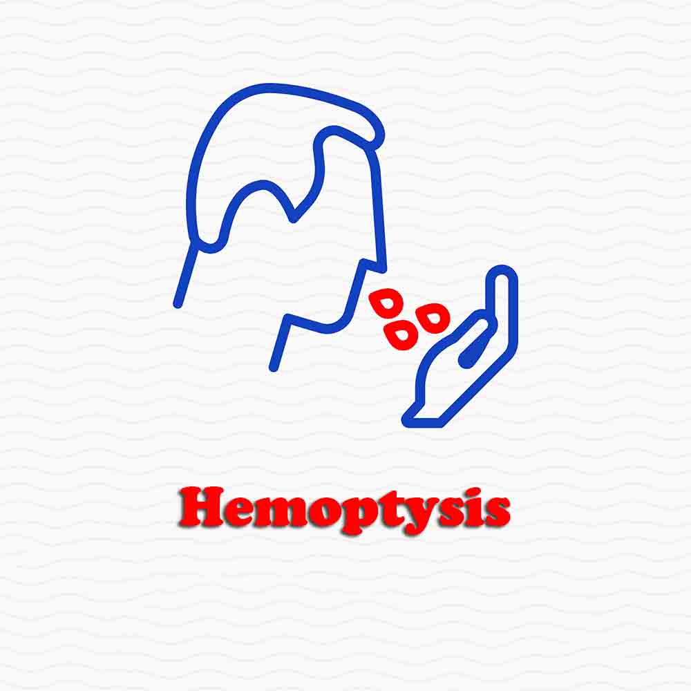

Sixth, hemoptysis, which is a severe sign that potentially indicates an underlying lung infarction due to a complete cessation of blood to lung segment that leads to tissue hypoperfusion and the death of tissues supplied by the obstructed vessel know as wedge lung infarction. This is not a common sign in pulmonary embolism, but a life-threatening problem that needs urgent medical attention.
7. Hypotension

Seventh, hypotension (i.e., low blood pressure), a severe sign in the context of a pulmonary embolism that denotes the need for an aggressive treatment strategy and close monitoring as it could be the beginning of a catastrophic complication known as obstructive shock. Hypotension is usually due to abrupt obstruction of the normal central circulation of blood from the right side of the heart through the lung for oxygenation and then back carrying oxygenated blood to the left side of the heart. When this is altered it causes low cardiac output state and hypotension.
8. Shock

Eighth, the patient may enter a Shock state, which is a circulatory failure to provide tissues with enough oxygen and nutrients to meet their metabolic needs. This life-threatening state may present as an abnormal level of consciousness due to decreased cardiac output and will manifest as unrecorded or severely low blood pressure values, acute kidney injury, and multi-organ failure.
9. The accompanying symptoms


Ninth, a group of non-specific manifestations including fever, syncope, abdominal pain, productive cough, wheezes, arrhythmia, atrial fibrillation or flutter and delirium may be the precipitating presentation or the accompanying symptoms that make the patient look for medical consultation in the emergency room or clinic. Also, the manifestations of the underlying cause may be the primary complain like lower limb swelling, pain, or hotness in the case of deep vein thrombosis.
The wide range of symptoms and their variability and non-specific nature plus the different stages of the severity of the disease from mild and chronic forms to acute life-threatening conditions make the diagnosis of Pulmonary embolism challenging and some times difficult, requiring the opinion of an expert Pulmonologist or Cardiologist or a complete medical team to confirm the diagnosis.
Many clinical score systems are established to assess the patient in order to improve the susceptibility of the diagnosis, as in the Well’s score. Clinical data collected from the patient is not enough to confirm the diagnosis as Pulmonary thromboembolism is a radiological finding and hence the use of Multi-slice CT pulmonary angiography will be needed in most of the cases to confirm or exclude the diagnosis and may help to guide the medical team to rule out any other diagnosis.
Pulmonary Embolism Causes
Pulmonary embolism is defined as an obstruction of the pulmonary artery by an embolus, i.e. blood clot, that originates in deep veins of the lower limbs or pelvis, and then a part of it is detached and lodged in one of the pulmonary arteries. When it is big enough, it may obstruct the main pulmonary artery, causing a critical condition known as massive pulmonary embolism that leads to hemodynamic compromise. Clearly, the condition always starts with deep vein thrombosis (DVT), and then complicates with pulmonary embolization.
In susceptible persons with several risk factors, platelet adhesion and aggregation take place forming a platelet nidus in the veins of the lower limbs or pelvis. This may also happen to a lesser extent in the upper limb. This event precedes thrombosis, increasing platelet aggregation and recruiting a fibrin network that filters more platelets from the blood, leading to a progressive stagnation and more thrombosis. Ultimately, a whole thrombus is formed. Blood fibrinolytic substances interacts with the thrombus, and then leads to partial dissolution. Detached particles of this thrombus will form the embolizing pulmonary thrombi that obstruct the pulmonary vessels.
So, why does venous thromboembolism take place?
Many acquired and congenitally inherited risk factors influence blood coagulability. For instance, thrombophilia is defined as hypercoagulable conditions that predispose people to an increased risk of venous thrombosis. These conditions are classified into acquired and inherited.
The most common inherited risk factors that are incorporated in venous thromboembolism are as follows:
-
- Factor V Leiden
- Prothrombin gene mutation G20210A
- Protein C and S, i.e. natural anticoagulants, deficiency
- Increased blood homocysteine
- Antithrombin
- Elevated factor VIII levels
On the other hand, the most common acquired risk factors are:
-
- Prolonged immobilization, long travel trips more than 3 hours in susceptible individuals
- Trauma
- Pregnancy
- Oral contraceptive pills
- Hormonal replacement therapy
- Malignancy
- Antiphospholipid syndrome (Lupus anticoagulant/ anticardiolipin)
- Heparin induced thrombocytopenia
- Inflammatory bowel disorders
- Central venous catheters
- Pacemakers
- Nephrotic syndrome
- Dehydration and increased blood viscosity
- Disseminated intravascular thrombosis
Hypercoagulability screening should be considered in patients with:
-
- Idiopathic Venous thromboembolism,
- Positive family history of venous thromboembolism,
- Early adulthood venous thromboembolism, i.e. first thrombotic event before the age of 50 years,
- thrombosis at unusual locations,
- Resistance to anticoagulation i.e. patients who experience thrombotic attacks while being properly anticoagulated medically,
- Recurrent thrombotic attacks.
Diagnosis of Pulmonary Embolism


The cornerstone of confirmation or exclusion in the diagnosis of pulmonary thromboembolism is imaging modalities specific for deep vein thrombosis and pulmonary embolism, which include:
I. Deep vein thrombosis
1. Venography
It is the method of choice to diagnose and visualize the extension of deep vein thrombosis. Now, it is rarely performed because of its invasive nature and the presence of accurate non-invasive modalities.
2. Duplex ultrasonography
It is currently the standard method for initial screening and diagnosis of deep vein thrombosis. By being reliable, non-invasive, of wide variability and easy interpretation, it gained its current position in the workup, and it is routinely ordered for all patients admitted to the ER with query deep vein thrombosis.
3. Magnetic resonance imaging
Rarely ordered for this purpose and with a limited value.
II. Imaging specific for pulmonary embolism:
1. CT pulmonary angiography
It is now considered to be the gold standard for diagnosis and risk stratification of pulmonary embolism, as it has a very high sensitivity and specificity. CT pulmonary angiography localizes the thrombus and its extension, and can be used for follow up and to exclude other mediastinal and parenchymal causes. It is also very helpful to confirm presence of lung infarction.
2. Ventilation – perfusion scanning (V/Q)
An important modality in the diagnosis of pulmonary embolism by showing segmental hypoperfusion. It could be used when CT pulmonary angiography is not available or contraindicated due to renal causes.
3. Magnetic resonance imaging
May be used if CT is not available or contra indicated due to renal causes. If gadolinium enhancement, i.e. contrast, is used, its specificity and sensitivity will be significantly higher.
4. Echocardiography
It is of a limited role due to its low sensitivity and specificity, though it is done in almost all cases in order to exclude other cardiac causes of chest pain or dyspnea. Its sensitivity and specificity are about 59% and 77%.
Sometimes it can visualize a central pulmonary artery thrombus and this is a highly positive value.
From all the above, it is clearly obvious that the diagnosis of pulmonary embolism in a step-wise manner resembles a sort of puzzle. Doctors use the approach described above in most hospitals worldwide. It is not wise to start with higher imaging techniques to exclude the presence of the disease due to cost-effectiveness causes and to limit side effects of contrast, minimize waiting lists in emergency rooms and avoid of unnecessary admissions.
Pulmonary Embolism Treatment


The main goal of treating pulmonary thromboembolism is avoiding a life-threatening outcome by stopping the formation of blood clots and the progression of the disease. This reduces the chances of new episodes after the acute cardiovascular event that would increase the risk of death due to pulmonary thromboembolism. Another goal is to prevent the progression of pulmonary thromboembolism to chronic venous insufficiency, pulmonary hypertension, and other complications.
To achieve these goals, your doctor will likely follow these steps:
1. Treating your risk factors
There are risk factors for blood clot formation that can be addressed to reduce the incidence and complications of pulmonary embolism. For instance, your doctor might take out certain medications that will contribute to clot formation, such as oral contraceptives and vitamin K. Another measure will be controlling your weight, reducing the time you spent sitting, lying down or immobilized, lowering your blood pressure in cases of hypertension, and advising against tobacco use. In some cases, your doctor might notice a blood imbalance of a substance called homocysteine. When it’s elevated, he will prescribe folic acid with or without vitamin B6.
2. Early deambulation
It means walking or moving around closely after being treated for a pulmonary embolism. It is recommended in most cases unless the patient has a case of massive pulmonary thromboembolism with a large blood clot or several blood clots compromising various parts of the vascular network. It is also not recommended in cases of unstable blood clots and hypotension. Early deambulation or walking is recommended 5 to 7 days after starting treatment with anticoagulants.
3. Heparin
It is a potent anticoagulant administered subcutaneously or in intravenous solutions with strict control of laboratory parameters. Your doctor will order several exams and keep ordering them as you go through heparin treatment, which is why it requires to be hospitalized in order to be administered.
This drug inactivates the enzymes and blood factors that trigger blood clotting, and the type of heparin that is advised for pulmonary thromboembolism is low molecular weight heparin, which is equally effective as another group called unfractionated heparin. Doses are calculated considering the patient’s weight, and it is administered every 12 hours along with oral anticoagulants for 5 days or more.
∗ Heparin use should be closely monitored in a hospitalized patient.